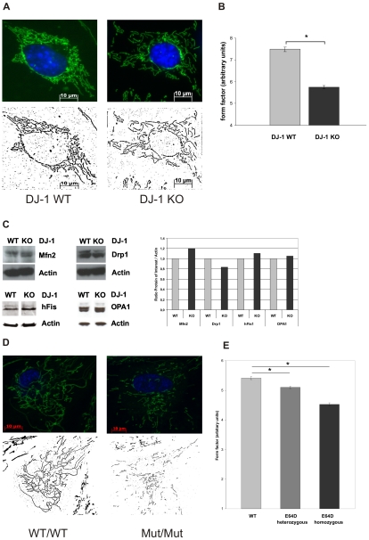
Figure 3. Effect of DJ-1 on mitochondrial morphology.
Mitochondrial morphology in living DJ-1 KO and DJ-1 WT MEF and in human fibroblasts from carriers of the E64D mutation in the DJ-1 gene and a healthy control were analyzed by life cell imaging microscopy (Cell Observer Z1, Zeiss, Germany) at 37°C using ApoTome®optical slides with 0.35–0.40 z-stacks. Mitochondria were stained with 200 nM MitoTracker® green FM (Invitrogen, USA), a specific mitochondrial dye, for 15 min at 37°C, nuclei were stained with Hoechst 33342 (Molecular Probes, USA; blue). (A) Fluorescence microscopy images of single mitochondria were analyzed using Image J 1.41o software (Wayne Rasband; National Institutes of Health, USA) for area, perimeter, major and minor axes. On the basis of these parameters, the aspect ratio (AR) of a mitochondrion (AR; ratio between the major and the minor axes of the ellipse equivalent to the object) and its form factor (FF; perimeter2/4π*area), consistent with the degree of branching, were calculated. (B) Mitochondrial branching as indicated by the form factor (FF) was significantly reduced in DJ-1 KO cells compared to WT (*p<0.001, Student's t-test). No significant differences in the AR were observed between DJ-1 KO cells and controls (not shown). Images from 45 individual cells were analyzed on three separate occasions by an investigator blinded to the experimental design. (C) Influence of DJ-1 on the expression of mitochondrial fission and fusion regulating proteins using WB analysis with specific antibodies against hFis1, OPA1, Drp1, and Mfn2. As a control for equal loading a specific antibody against β-actin was used. Densitometric quantification revealed no significant differences in the respective protein levels between WT and KO MEF. (D) Fluorescence microscopy images of single mitochondria of human fibroblasts from the E64D family and a healthy control (see supplemental figure S1) were analyzed using Image J 1.41o software (Wayne Rasband; National Institutes of Health, USA) as described previously. On the basis of these parameters, the aspect ratio (AR) of a mitochondrion (AR; ratio between the major and the minor axes of the ellipse equivalent to the object) and its form factor (FF; perimeter2/4π*area), consistent with the degree of branching, were calculated. (E) Mitochondrial branching as indicated by the form factor (FF) was significantly reduced in cells from the index patient carrying the homozygous E64D mutation compared to the healthy control (*p<0.001, Student's t-test). Also heterozygous carriers of the E64D mutation revealed a decreased form factor compared to control (*p<0.05, Student's t-test). Images from 40 individual cells of each proband were analyzed on three separate occasions by an investigator blinded to the experimental design.

