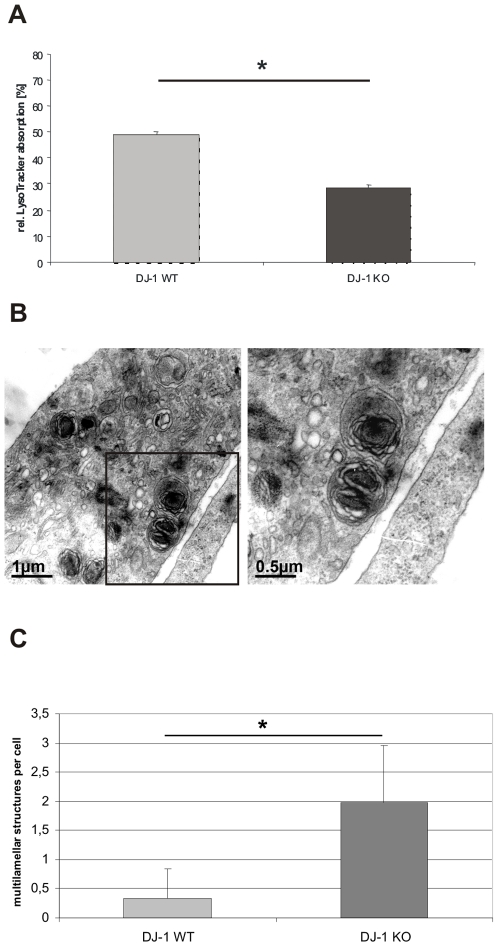
Figure 4. Lysosomal phenotype in DJ-1 WT and DJ-1 deficient MEF.
(A) Lysosomal activity was determined by FACS analysis. Cells were stained with the specific lysosomal dye LysoTracker® (Invitrogen, USA) 1 µM for 15 min at 37°C. The absorption of LysoTracker® was measured by FACS. The uptake of LysoTracker® is an indicator of the Lysosomal mass. DJ-1 KO cells revealed a significantly reduced uptake of LysoTracker® compared to WT MEF (*p<0.0001, Student's t-test). The experiments were performed in triplicate on three different days, the diagram represents 1 of 3 sets of experiments with similar results. (B) Ultrastructural analyzes of DJ-1 deficient MEF and MEF from DJ-1 WT littermates were performed by electron microscopy. MEF lacking DJ-1 show multilamellar structures reminiscent of modified lysosomes. Bars 1 µm or 0,5 µm, respectively. (C) Quantification of lysosomal-like structures in DJ-1 WT and DJ-1 KO MEF. Multilamellar structured lysosomes were counted per cell and were significantly more frequent in DJ-1 KO cells compared to WT controls (p<0.001, Student's t-test). A total of 150 individual cells were evaluated for the presence of multilamellar structures.

