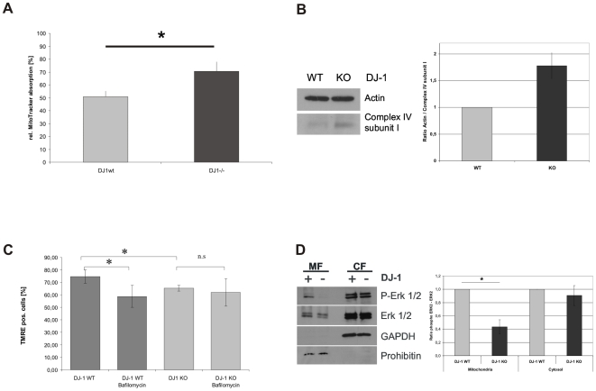
Figure 8. Influence of lysosomal degradation of mitochondria on mitochondrial mass and its mechanistic backround.
(A) To analyze mitochondrial mass in DJ-1 KO and DJ-1 WT MEF, cells were treated with 1 µM MitoTracker® green FM (Invitrogen, USA) for 15 min at 37°C. Absorption of MitoTracker® green FM was determined by FACS analysis. The uptake of MitoTracker® was used as an indicator for the mitochondrial mass. We found evidence for increased mitochondrial mass in DJ-1 KO cells compared to controls (*p<0.001, Student's t-test). (B) To further validate our observations on mitochondrial mass, we used an independent method quantifying complex IV subunit I as a mitochondrially encoded protein of the respiratory chain. We found increased protein levels of complex IV subunit I in KO cells compared to controls as indicative of an increased mitochondrial mass in the KO condition. (C) The influence of disturbed lysosomal activity on mitochondrial function was analyzed by measuring the MMP after treatment with BafA1 as an inhibitor of lysosomal function. Cells were treated overnight with 200 nM BafA1 (Calbiochem, Germany) and the integrity of MMP was determined by measuring the uptake of TMRE (Invitrogen, USA) by FACS analysis (CyAnADP, Beckman Coulter). (C) To analyze candidate pathways for DJ-1-mediated modulation of lysosomal degradation we investigated p42/p44 MAPK (ERK1/2) phosphorylation in the mitochondrial (MF) and cytosolic fraction (CF) of DJ-1 WT and DJ-1 KO MEF. Phosphorylation and expression of the ERK1/2 kinase was analyzed by WB using antibodies against phospho-ERK1/2 (Thr202/Tyr204). To define the fractions we used antibodies against prohibitin or GAPDH, as markers for the mitochondrial and the cytosolic fraction, respectively. Quantification of relative protein levels was performed by Image J software. The results indicate a significantly reduced level of phospho-ERK2 in the mitochondrial fraction (p<0.05; Student's t test).

