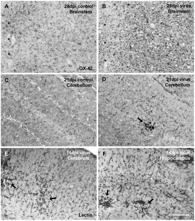
Fig. 3.
Besides the spinal cord, brainstem is another virus infection-sensitive brain area displaying evident microglial activation (A, B), asterisks indicating the midline. Microglial activation is generally less pronounced in the cerebral cortex, cerebellum, and other brain areas. However, clusters of activated microglia can be occasionally encountered in these areas, as exampled in virus-infected cerebellum (D), cerebral cortex (E), and hippocampus (F). (C) Cerebellum of a control animal at 21 dpi shows no microglial activation. (A-D) Microglia are immunolabeled with OX-42. (E, F) Microglia are labeled by lectin histochemistry. Scale bar, 200 μm.

