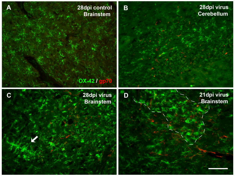
Fig. 4.
Double immunofluorescent staining for microglia (OX-42) and viral gp70 reveals lack of direct correlation between presence of activated microglia and PVC-211-infected endothelial cells. (A) Absence of gp70 immunoreactivity in the brainstem of a control rat. (B) A virus-infected cerebellum with virus infection but no microglial activation. (C, D) Virus-infected brainstems, with universal gp70 labeling while activated microglia in confined areas. (C) Activated microglia are spread diffusely throughout the field, with some aggregates (arrow), but do not accumulate around gp70-positive blood vessels. (D) Both resting and activated microglia (area of activation delineated by dashed line) are colocalized with widespread gp70 immunoreactivity in microvessels. Scale bar, 100 μm.

