Fig. 6.
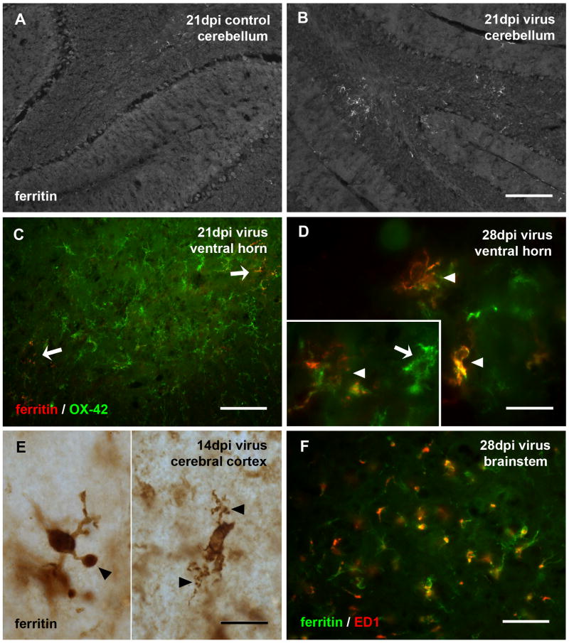
Expression of ferritin is increased following viral infection and associated with dystrophic microglia. (A, B) Low power view of the cerebellum in control and infected rats shows increased ferritin staining of microglia in the cerebellum. Ferritin-positive microglia in virus-infected spinal cord shows them to be a small subset of OX-42-positive cells (C, pointed by arrows). Double-labeling with anti-ferritin and OX-42 in spinal cord ventral horn (D) or single immunolabeling by ferritin in cerebral cortex (E, pieced together from two views) reveals them to exhibit microglial dystrophy evident as short, tortuous and fragmented cytoplasmic processes (arrowheads in D-E, and inset in D). The inset in D shows a hypertrophic OX-42-positive cell that is negative for ferritin (arrow). Double-labeling with anti-ferritin and ED1 in the brainstem (F) shows ED1 immunoreactivity present in some ferritin-positive microglia. Scale bar, 200 μm in B for A and B, 100 μm in C, 20 μm in D-E and inset, 50 μm in F.

