Fig. 7.
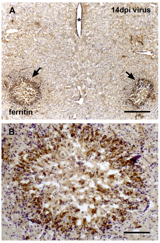
Selective degeneration of the red nuclei in midbrain is associated with intense ferritin immunoreactivity. (A) Localized increases in ferritin staining are evident in red nucleus areas bilaterally (arrows), aterisks indicating the cerebral aqueduct. (B) High magnification reveals necrosis and loss of rubrospinal neurons. The intensely immunoreactive perimeter is characterized by hypercellularity, and immunorecative cells likely represent degenerating microglia, cresyl violet counterstain. Scale bar, 400 μm in A, 100 μm in B.

