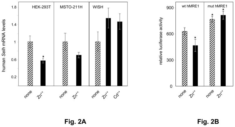
Fig. 2. Human Selh expression is regulated by metals.
A. Human Selh mRNA expression was assessed in HEK-293T, MSTO-211H, and WISH cell lines in the absence (striped bars) or presence (black bars) of heavy metal treatment, as described in the text and in Table 1. mRNA levels relative to Hprt were analyzed by real time PCR (n=3) and are plotted as mean ± SD from 3 experiments. Student’s paired t test was used for statistical evaluation; (*) indicates P<0.05. B. Wt and mut human MRE1 luciferase expression vectors introduced into HEK-293T cells were used to evaluate Selh promoter activity in the absence (striped bars) or presence (black bars) of Zn++. Relative luciferase units are represented as ratio of firefly luciferase activity driven by Selh promoter fragment to Renilla luciferase activity from co-transfected control plasmid (pRLSV40). Each value represents the mean ± standard deviation of four independent transfection experiments, each performed in triplicate. Asterisks indicate values below the nominal P<0.05 compared to untreated wt MRE1 construct.

