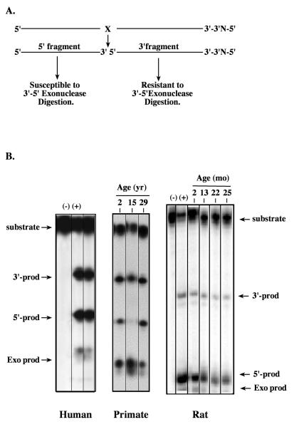
Figure 1. Exonuclease activity in extracts prepared from rodent, primate and human brain tissue.
Tissue homogenates of freshly frozen cortical tissue from 2 month to 25 month-old male rats, 2 yr, 15 yr and 29 yr old male rhesus monkeys (Maccaca mulatta) and a 77 yr male (PMI 2.25h) were prepared according to previously published methods (Kisby et al., 1997). A. An aliquot of each extract (0.1 μg protein) was incubated at 37°C for 10 min with a double-stranded 5′-5′ oligonucleotide substrate (50 fmol) containing a tetrahydrofuran site that had been end labeled with 32P-ATP (Muniz et al., 2008). B. Representative gels showing bands for both APE activity (i.e., 5′ and 3′ cleavage products) and exonuclease activity (exo product). (+) Purified E. coli endonuclease IV; (−) no endonuclease IV.

