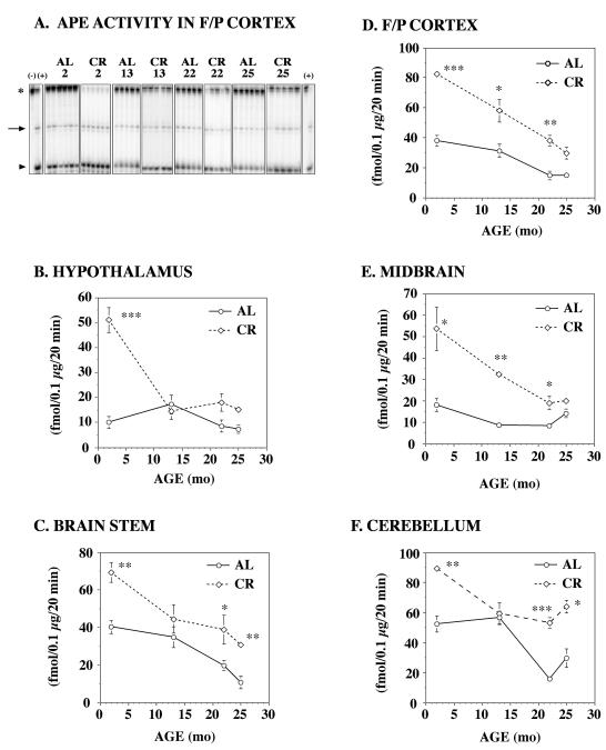
Figure 3. AP endonuclease (APE) activity in various brain regions of aging CR rats.
Homogenates of freshly frozen brain tissue from 2mo, 13 mo, 22 mo, and 25 mo F344 rats on an ab libitum (AL) or caloric restricted (CR) diet (n=6/age group) were prepared as previously described by Kisby et al. (1997) and examined for APE activity using the 5′-5′ oligonucleotide probe (50 fmol) described in Fig 1. A. A representative autoradiogram showing AP endonuclease activity in the F/P cortex of rats on an AL or CR diet. The arrow denotes the 3′ cleavage product, the star denotes the substrate and the arrowhead denotes the 5′ cleavage product. Negative control (lane 1) and positive control (2nd and last lanes). AP endonuclease activity was measured in the hypothalamus (B), brain stem (C), frontal/parietal (F/P) cortex (D), midbrain (E) and cerebellum (F). In general, APE activity declined with age, but CR significantly retarded the rate of decline. Significantly different from AL (*p<0.05, ** p < 0.01, *** p <0.001).

