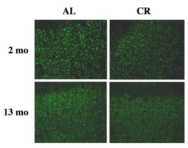
Figure 5. Distribution of APE in the frontal/parietal cortex of aging CR rats.
Photomicrographs of representative sections from the frontal/parietal cortex of 2 mo and 13 mo AL and CR rats probed with an antibody to APE. Note that the distribution of immunopositive cells is uniform in AL and CR rats. Magnification 40×.

