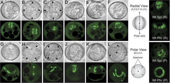
Fig. 5.
Mitotic and cytokinetic apparatus in TMBP200 RNAi lines. Spindles (A–F) and phragmoplasts (G–L) from microspores undergoing aberrant mitosis or cytokinesis were viewed with DIC (upper panel) and with CLSM (lower panel). Images are shown in radial view (A, D, E, F, G, J, K), GP view (B, C, H, I) or at an angle in between (L). Arrowheads (B, C, H, I, L) indicate positions of pollen apertures. Mutant spindles represent bipolar spindles 90° rotated (A, B, C) and multipolar spindles (D, E, F). Mutant phragmoplasts represent single but misoriented (G, H) and complex branched or fragmented forms (I, J, K, L). An orientation schematic and wild-type spindle and phragmoplast images are shown for radial (R) and polar (P) views.

