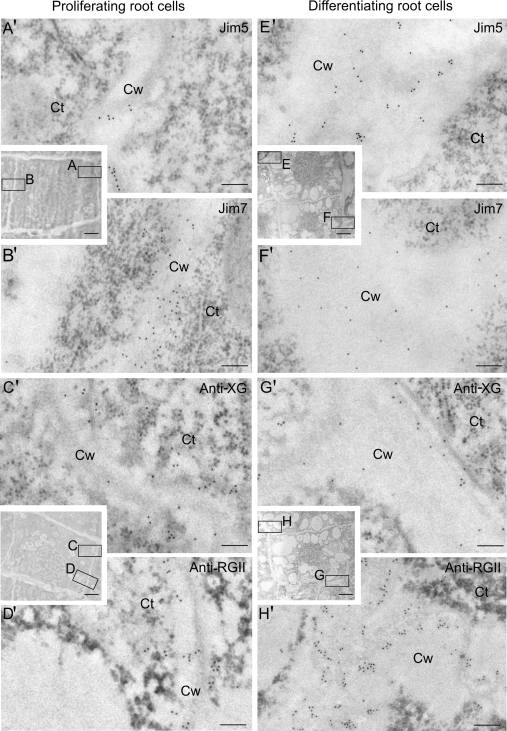
Fig. 3.
Immunogold labelling of cell wall antigens on proliferating (A–D, A′–D′) and differentiating (E–H, E′–H′) cells of the root apical meristems of Allium cepa L. Electron micrographs of Lowicryl ultrathin sections. Inserts: low magnification micrographs showing the structural organization of the proliferating and differentiating cells of the root apex. Location and distribution at the ultrastructural level of JIM5, JIM7, anti-XG, and anti-RGII antigens in the newly-formed walls (squares A, B, C, D) of the proliferating area (A′, B′, C′, D′) and in the more developed walls (squares E, F, G, H) of the differentiating cells (E′, F′, G′, H′) of the elongation area. Ct, cytoplasm; Cw, cell wall. Bars: (A′, B′, C′, D′, E′, F′, G′, H′) 200 nm; inserts: 5 μm.

