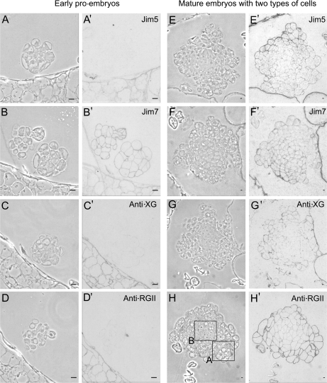
Fig. 5.
Immunolocalization of cell wall antigens on pollen-derived embryos of Capsicum annuum L. during two developmental stages: early pro-embryos (A–D, A′–D′) and more developed embryos with two cell types (E–H, E′–H′). Immunogold labelling and amplification with silver enhancement with antibodies JIM5, JIM7, anti-XG, and anti-RGII observed under phase contrast (A–H), to visualize the general structure, and bright field (A′–H′) to visualize the immunoreaction. The two cell types of the more developed embryos are indicated by the squares in (H); differentiating cells in square A, and proliferating cells in square B. Bars: (A, B, C, D) 10 μm; (E, F, G, H) 50 μm.

