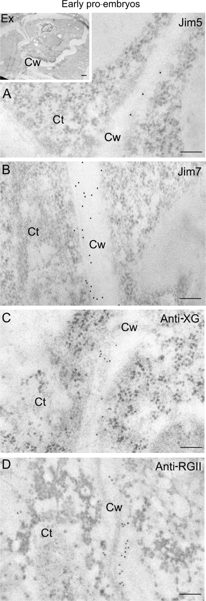
Fig. 6.
Immunogold labelling of cell wall antigens in early pollen-derived proembryos of Capsicum annuum L. Electron micrographs of Lowicryl ultra-thin sections. Insert: low magnification micrograph showing proliferating cells of an early proembryo still surrounded by the special pollen wall, the exine (Ex). (A, B, C, D) High magnification micrographs showing the immunogold labelling with JIM5 (A), JIM7 (B), anti-XG (C), and anti-RGII (D) in the cell walls (Cw). Ct, cytoplasm. Bars: (A, B, C, D) 200 nm; insert: 1 μm.

