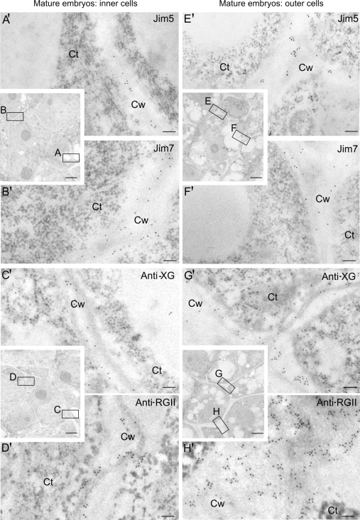
Fig. 7.
Immunogold labelling of cell wall antigens in the two cell types of late-mature pollen-derived embryos of Capsicum annuum L. Electron micrographs of Lowicryl ultrathin sections. Inserts: low magnification micrographs showing the structural organization of the cells of the inner and outer areas of the mature embryo. Location and distribution at the ultrastructural level of JIM5, JIM7, anti-XG, and anti-RGII antigens in the newly-formed walls (squares A, B, C, D) of the inner proliferating cells (A′, B′, C′, D′) and in the more developed walls (squares E, F, G, H) of the outer differentiating cells (E′, F′, G′, H′) of the mature embryo. Ct, cytoplasm; Cw, cell wall. Bars: (A′-H′) 200 nm; inserts: 1 μm.

