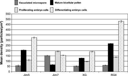
Fig. 8.
Quantification of immunogold labelling of cell wall antigens during pollen development and in pollen-derived embryos of Capsicum annuum L. The histogram shows the labelling density as the number of gold particles per area unit (square micrometre). Striped and black bars correspond to pollen development: striped bars correspond to vacuolated microspores, black bars to mature pollen. Grey and dotted bars correspond to pollen-derived embryos: grey bars correspond to proliferating cells of early pro-embryos, dotted bars to differentiating cells of mature embryos.

