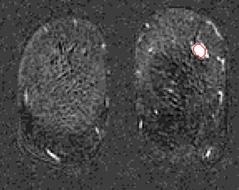
Figure 3b:
Coronal MR images show segmentation (red outline) of (a) IDC lesion with positive LNs in 34-year-old woman, (b) IDC lesion with negative LNs in 39-year-old woman, (c) DCIS lesion in 66-year-old woman, and (d) benign lesion in 48-year-old woman. (e) Corresponding characteristic kinetic curves for these four breast lesions.

