
Figure 1d:
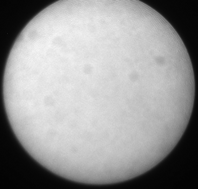
(a) A dual-port optical imager, which can be positioned percutaneously by using a guiding needle, has an input to deliver excitation light into the target and an output to detect emission light from the target. (b) With real-time ultrasonographic (US) imaging guidance, the optical imager (arrowheads) is percutaneously inserted to image a target artery (arrow) that received gene transfer for expressing green fluorescent protein. (c, d) Direct views from percutaneous optical imaging show that the emission light is brighter in (d) the green fluorescent protein–treated artery than in (c) the saline-treated control artery. (e, f) Results of fluorescent microscopy confirm higher green fluorescence from both the intima (arrows in f) and media (M) of (f) the green fluorescent protein–treated artery compared with (e) the autofluorescence from the internal elastic lamina (arrows) of the saline-treated control artery. (Reprinted, with permission, from reference 4.)