
Figure 1.
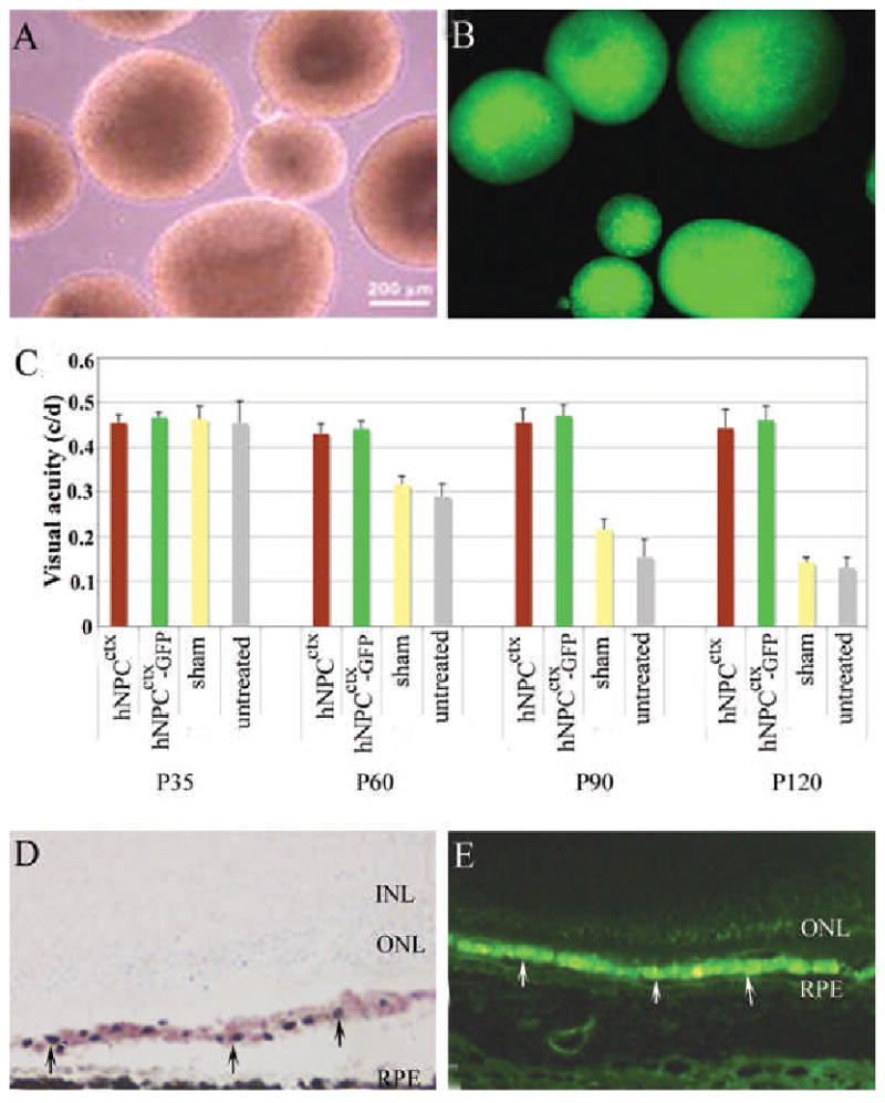
A: Light microscopy of human prenatal cortical progenitor cells (hNPCctx) cultured as neurospheres; B: fluorescence microscopy of GFP transduced hNPCctx; C: Visual thresholds from native hNPCctx, prelabeled hNPCctx-GFP, sham and untreated eyes from RCS rats (n=10/group) tested at four time points. There is no difference between native hNPCctx and hNPCctx-GFP treated groups (p>0.05); however, significantly different between cell-injected and shame/untreated controls (p<0.01). Two weeks after subretinal injection of hNPCctx, the distribution of donor cells (arrowed) is similar, as shown in D (retinal cross section with donor cells stained with human nuclear marker on cresyl violet background) and E, (hNPCctx –GFP shown green). No donor cells were seen in the sensory retina at any time.