
Figure 3.
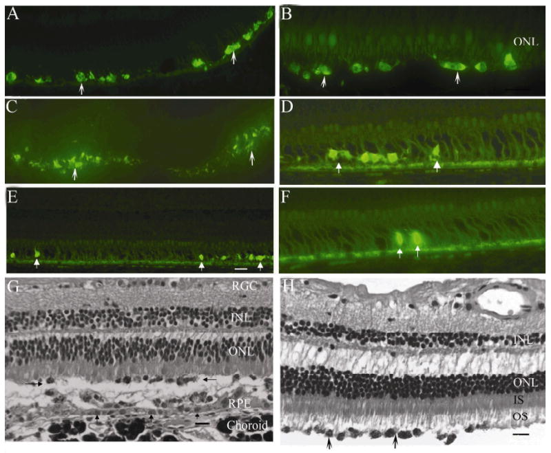
Immunofluorescence images of hNPCctx-GFP cells injected into the subretinal space of primate eyes at day 3 (A), 7 (B &G), 14 (C) and 31 (D, E, F &H) after injection (monkeys 1, 2, 3 and 4, respectively). G and H are black and white images of cresyl violet stained sections at day 7 (monkey 2) and day 31 (monkey 4) after injection, respectively. hNPCctx-GFP (white arrows in A-F, black arrows in H) were distributed in the subretinal space and in contact with host outer segments. G: donor cells are indicated by left and right pointing arrows, RPE layer by vertical arrows; scale bars equal 20μm). H: the process of tissue fixation has resulted in artefactual detachment of the RPE layer and enlargement of spaces between inner and outer nuclear layers. At seven days after injection, photoreceptor outer segments were shortened (B & G) but by day 31 (H), outer segments were well formed and donor cells appeared closely apposed.