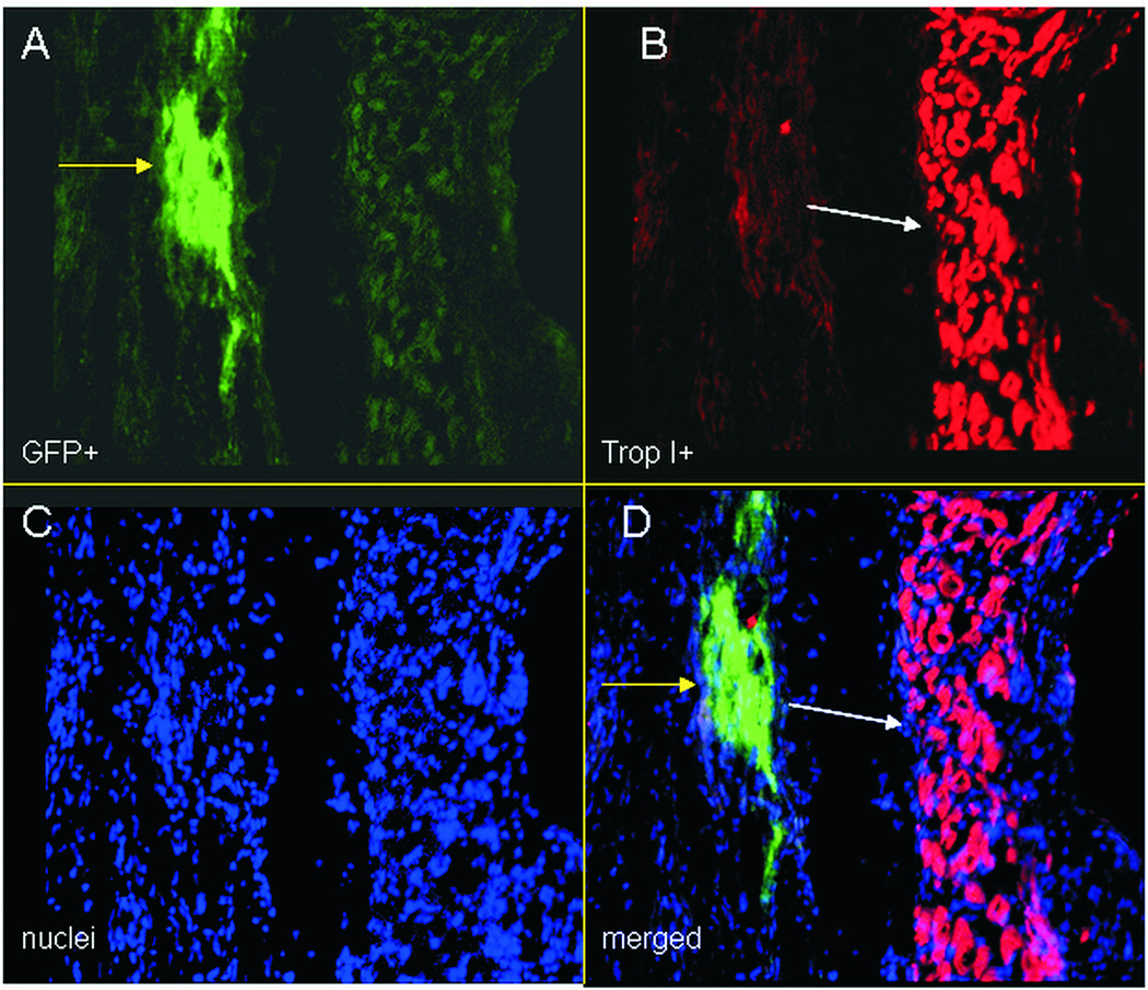
Figure 1.
Immunocytochemistry images of eGFP overexpressing cardiac derived stem cells transplanted in the hearts of infarcted rats, at 21 days post cell injection. A: eGFP+ cells (green-yellow arrow), B: cardiac Troponin I+ cells (cardiomyocytes-red-white arrow), at the infarct border zone, C: nuclei (stained with Hoechst 33342-blue), d: merged image. eGFP does not co-localize with cardiac TropI, indicating that cells have not differentiated yet.

