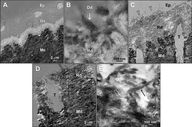
Figure 4.
TEMs taken from unstained, non-demineralized sections of acid-etched dentin in the Portland cement-based biomimetic remineralization control group. (A) A specimen retrieved after 2 mos of biomimetic remineralization in the polyacrylic acid- and PVPA-containing SBF revealed discrete islands of partial remineralization (open arrowheads) and a 3-µm-thick zone of partial remineralization (pointer) over the original demineralization front. (B) A high-magnification view of partially remineralized dentin depicted in Fig. 4A. Collagen fibrils along the dentin surface (arrow) were heavily remineralized with nanocrystals. Individual nanocrystals (open arrowhead) could also be seen in the less-highly-remineralized regions. (C) A 3-month-old specimen retrieved from the polyacrylic acid- and PVPA-containing SBF revealed heavier remineralization than extended from the dentin surface to the mineralized dentin base. Remineralization was absent or sparse in some locations (between open arrowheads). (D) A 4-month-old specimen retrieved from the polyacrylic acid- and PVPA-containing SBF showing more extensive but incomplete remineralization of the acid-etched dentin. The original demineralization front was difficult to be discerned (arrow). (E) A high-magnification view of Fig. 4D. Intrafibrillar remineralization of a collagen fibril could be seen in an incompletely remineralized region (arrow). Generic abbreviations: Dd, demineralized dentin; Rd, remineralized dentin; Md, naturally mineralized dentin; Ep, epoxy resin; T, dentinal tubule.

