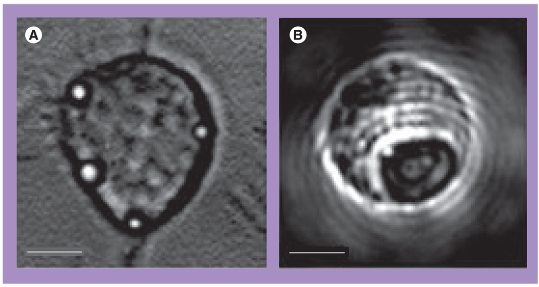
Figure 11. Nanoparticle-generated photothermal bubbles in cells.
(A) A549 (C225-positive) cell with clusters of nanoshells exposed to a single laser pulse (10 ns, 750 nm, 0.7 J/cm2). (B) K562 leukemia cell treated with 30-nm gold nanoparticles and a single laser pulse (10 ns, 532 nm, 1.7 J/cm2) has yielded a big photothermal bubble. Scale bar is 10 µm.
Reproduced with permission from [53].

