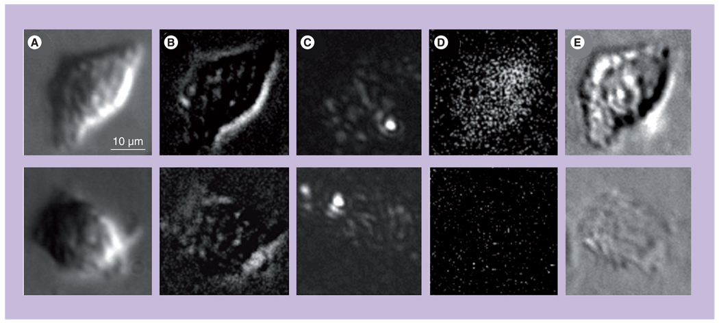
Figure 13. Images of the damaged (top panel) and surviving (bottom panel) A549 cells after their incubation with 170-nm gold nanoshells and the follow-up exposure to a single pump laser pulse that induced the photothermal bubbles.
(A) White light image prior to exposure to pump laser pulse. (B) Side scattering image prior to exposure to pump laser pulse shows nanoshell-related signals. (C) Side scattering image during exposure to pump laser pulse shows photothermal bubbles. (D) Fluorescent image after the exposure to pump laser pulse shows epidium bromide fluorescence. (E) Differential white light image shows changes in the cell after exposure to pump laser pulse.
Reproduced with permission from [38].

