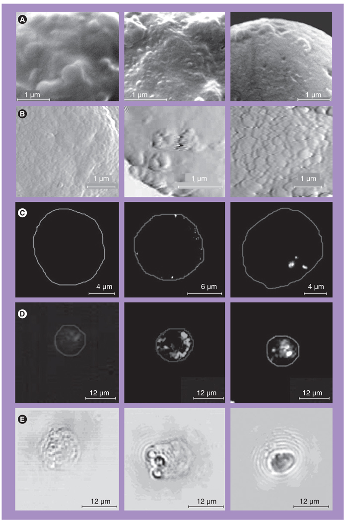
Figure 9. Images of individual K562 cells obtained before and after interaction with gold nanoparticles.
Left: control; center: after targeting at 4°C with 30-nm gold nanoparticles (NPs); right: after targeting at 37°C with 30-nm gold NPs. (A) Images obtained with scanning electron microscope. (B) Images of K562 cells obtained with atomic force microscope. (C) Images obtained with fluorescent microscope. (D) Images obtained with optical side scattering microscope. (E) Images obtained with time-resolved photothermal microscope show the photothermal bubbles: small ones in the center image and large one in the right image, while no photothermal bubbles were detected in the left image.
Reproduced with permission from [51].

