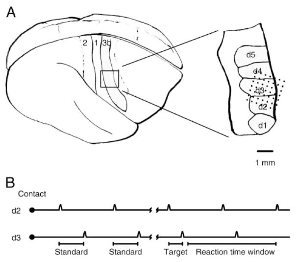
FIG. 1.
A: implant location. Left: side aspect of the owl monkey brain. Right: anterior. Somatosensory strip in area 3b was implanted. Inset: approximate position of the 49 microelectrode implants in animal 1. Electrodes were implanted across the 2nd (d2), 3rd (d3), and 4th (d4) digital representations on the left hand. B: behavioral task. A trial began with an orienting response, the animal initiating contact with the tips of 2 motors. Then, 2 to 6 standard tap pairs were repeated. After the tap interval changed to the 100-ms target, which was shorter than the standard interval (200 ms), the animal could remove its hand and receive a reward.

