Figure 1.

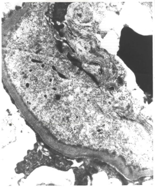
Electron-microscopy: Subendothelial electron-dense deposits were seen along the basement membrane. Mesangial electron-dense deposits were also detected. These histologic changes were consistent with MPGN, type 1.

Electron-microscopy: Subendothelial electron-dense deposits were seen along the basement membrane. Mesangial electron-dense deposits were also detected. These histologic changes were consistent with MPGN, type 1.