
Fig. 1.
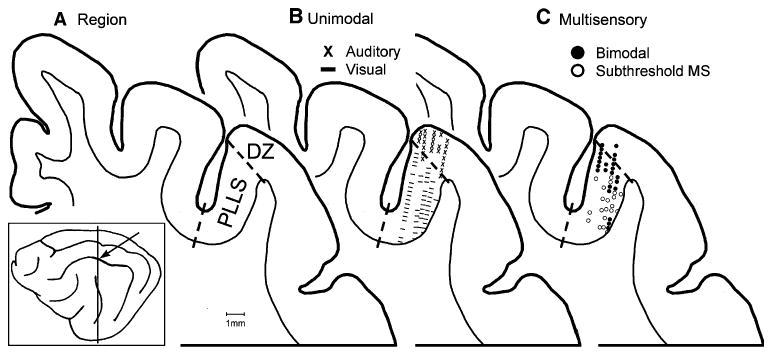
Sensory and multisensory processing in visual PLLS. The location of the posterolateral lateral suprasylvian (PLLS, arrow) visual area and the level of the coronal section (vertical line) are shown on the lateral view of the cat brain (box). Section A shows the location of the visual PLLS in the lateral bank and fundus of the suprasylvian sulcus and the auditory DZ at the lip of the sulcus. This same coronal section is used again in (B), and (C). In B the locations of physiologically identified visual PLLS neurons (dashes) and auditory DZ neurons (x's) are shown (from Allman and Meredith 2007). In C the location of physiologically recorded bimodal (closed circles) and subthreshold (open circles) multisensory neurons (1 marker = one or more neurons; see Allman and Meredith 2007) are shown. Note that bimodal neurons were clustered along the outer border of PLLS with DZ, while subthreshold multisensory neurons were found at deeper locations along the bank of the suprasylvian sulcus. PES posterior ectosylvian sulcus