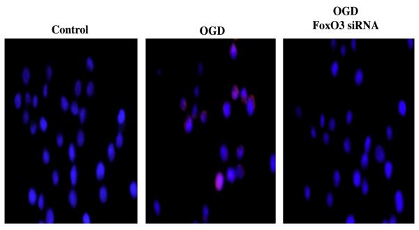
Figure 1. FoxO3a can control the activity of caspase 3.
Inflammatory microglial cells were exposed to oxidative stress through oxygen-glucose deprivation (OGD) and caspase 3 activation was determined six hours after OGD exposure through immunocytochemistry with antibodies against cleaved active caspase 3 (17 kDa). Representative images show no caspase 3 activity staining (blue) in control (untreated cells), but active caspase 3 staining (red) in cells following OGD. In contrast, gene silencing of FoxO3a during transfection with FoxO3a siRNA yields significantly reduced caspase 3 activity with demonstration of minimal red staining.

