
Figure 1.
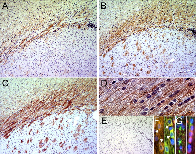
IHC detection of p-mTOR Ser 2448 in subcortical white matter during postnatal rat CNS development. A–C, Fixed, paraffin-embedded sections from postnatal day 14 (A), day 18 (B), or day 21 (C) were used for IHC staining of p-mTOR Ser 2448 (detected with vector-red). D, Image (40×) of p-mTOR staining in the WM at P21. E, Control d 21 section stained with secondary antibody alone. Sections were counter-stained with hematoxylin. F, 40× image of p-mTOR staining in the WM at d 21. Immunostaining of p-mTOR (TRITC), MBP (FITC), and DAPI in corpus callosum. Panels show two fields with colocalization of p-mTOR and MBP (arrowheads). G, Immunostaining of p-mTOR (TRITC), GFAP (FITC), and DAPI in corpus callosum. Panels show GFAP and p-mTOR in separate cells in two fields.