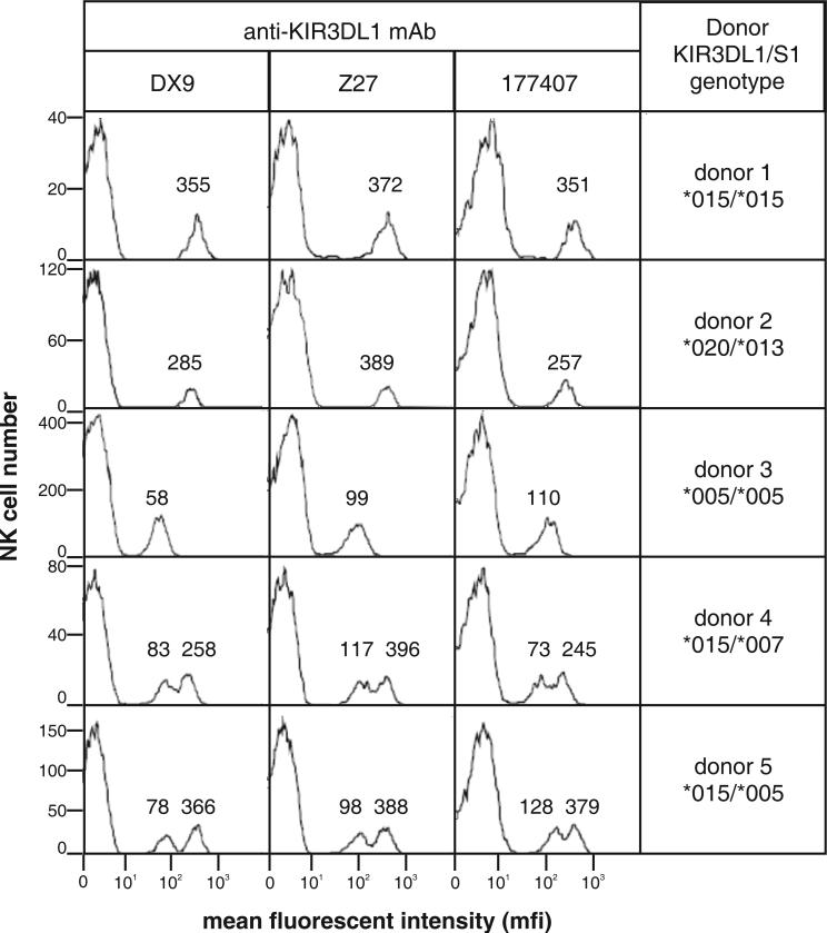
Figure 4. Like DX9 and Z27, the 177407 antibody distinguishes KIR3DL1 allotypes expressed on the surface of peripheral blood NK cells.
NK cells were isolated from the blood of donors of known KIR3DL1/S1 genotype and analyzed for expression of 3DL1/S1 using the DX9, Z27 and 177407 antibodies and flow cytometric analysis as described (11). KIR3DL1 allotypes are expressed at different levels on the NK cell surface and bind the DX9 and Z27 antibodies to high level (for example, 3DL1*015 and *020) or to low level (for example, 3DL1*005 and *007) Because minority subsets of NK cells express KIR3DL1, in heterozygous donors there are different subsets of NK cells expressing the two alleles (as well as a small number of cells expressing both alleles). If the donor is homozygous for a high-expressing allele (donor 1) then a unimodal distribution of 3DL1-expressing cells is observed, with a relatively high level of antibody bound. The value for the mean fluorescent intensity (mfi) of cells within each peak is given above the peak: 355 with DX9 of donor 1, for example. Similarly, a donor homozygous for a low-expressing allele (donor 3) gives a unimodal distribution with relatively low mfi. Heterozygote donors with a high- and a low-expressing allele (donors 4 and 5) give a bimodal distribution because of the two subsets of NK cells, one expressing the high allele and the other the low allele. The very low binding of 3DS1*013 to Z27 (26, 28) is seen in donor 2 as slight shoulder on the peak of KIR-negative NK cells (the majority) with mfi <1.

