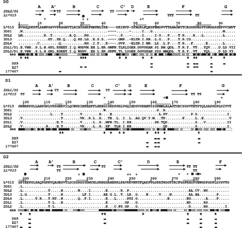
Figure 6. Multiple sequence alignment for the extracellular ligand-binding domains domains (D0, D1 and D2) of human KIR.
Included in the alignment were representative KIR of all four human lineages: 2DL4*003 of lineage I (Q99706); 3DL1*015 (P43529), 3DS1*013 (Q14943), and 3DL2*001 (P43630) of lineage II; 2DL1*001 (P43626) and 2DL2*001 (P43627) of lineage III, and KIR3DL3*001 (Q8N743) of lineage V. Note that 2DL4 has only D0 and D2 domains, but no D1 domain. Above the alignment are shown the elements of secondary structure in the KIR3DL1 model and the template used (2DL2 for D0 and 2DL1 for D1-D2). The arrows indicate positions of the β strands and T the residues implied in bulge conformation. Below the alignment: each amino acid is assessed to be exposed (black), buried (white) or intermediate (gray) according to their accessible solvent area (asa); large stars indicate the sites of mutation studied here, rectangles the mutations that gave a significant difference in antibody binding compared to 3DL1*015. Above the alignment: + denotes residues in the electropositive site predicted for D0 and D1; * indicates residues that contribute to the hydrophobic binding pocket; squares indicate residues involved in predicted salt bridges and hydrogen bonds; and gray lines show the position of loops that form the predicted HLA-binding site, or are close to it.

