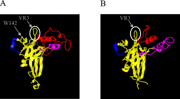
Figure 4.
Crystal structure of the receptor binding domain of the Friend murine leukemia virus (A) and FeLV-B (B). VRA, B and C loops are shown in red, purple and blue. VR3 loop is encircled. The pictures are generated using the Rasmol software and coordinates (A: 1AOL B: 1LCS) from the RCSB protein data bank http://www.rcsb.org/pdb/home/home.do.

