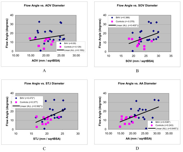
Figure 6.
Scatter diagrams showing distribution systolic blood flow jet angles of aortic outflow as a function of aortic diameters, corrected for BSA, and at the level of the aortic valve (A), sinus of Valsalva (B), sinotubular junction (C) and ascending aorta (D). Regression lines are shown of line fits with all subjects combined. An asterisk with the Pearson correlation coefficient in the figure legend indicates a statistically significant correlation.

