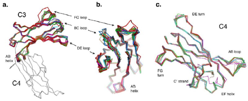
Figure 5.

(a) Side view of an overlay of the C3 domains aligned on each other, showing the variable FcεRIα-binding loops: BC, DE and FG. (b) C3 domain overlay rotated 90° from the view in (a). (c) An overlay of the C4 domains aligned on each other, showing the variable AB loop, the DE and FG turns, the C′ strand and EF helix.
