Figure 6.


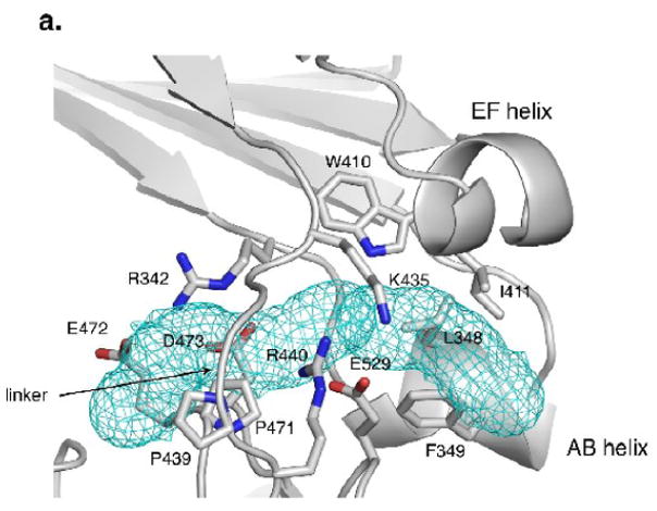
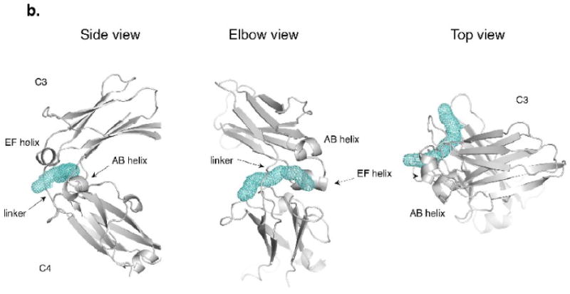
The elbow region at the linker between the C3 and C4 domains. (a) The hydrophobic pocket at the elbow region in the P21 chain D (PD). Hydrophobic residues W410, I411, L348, and F349 are joined by the ring of P471 as well as the aliphatic portion of R342 in forming the pocket. The entry to the pocket is partially covered by charged and polar residues, including K435 and E529 on the exterior face and R342 and D473 at the dimer interface side. The pocket continues into a tunnel that opens on to the dimer interface side. (b) The tunnel found by MOLE18 in the PD chain (side, elbow and top views). Additional residues contacting the tunnel in PD include T434, T436, D473, E472, R342, F503, M470 and P439.
