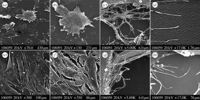
Figure 5.
Scanning electron micrographs of osteoprogenitor interaction with NSQ50 nanopits compared with planar control. On the NSQ50 topography (a–d) cells could adhere and differentiate to form a dense aggregate of cells similar in appearance to bone nodules (a,b). (c,d) Cells used their filopodia to sense nanopit topography surface. On the flat surface (e–h) cells were fully spread and proliferated to form a confluent tissue layer (e,f). Also, (g,h) cells using their filopodia to sense the substratum surface can be seen.

