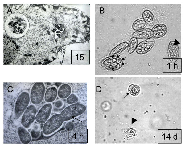
Figure 1.
A microscopic study of interactions between L. monocytogenes and T. pyriformis. A. Bacterial uptake by T. pyriformis in 15 minutes after the microorganisms were mixed. B. T. pyriformis cells in 1 h after the microorganisms were mixed. Multiple phagosomes within one cell are shown with arrows. T. pyriformis cell without phagosomes is shown with an arrowhead. C. Intraphagosomal bacteria. Dividing bacterium is shown with an arrow. D. Cysts (an arrow) and cell remnants (an arrowhead) after two weeks of incubation. The images were captured with transmission electron (A, C), or light (B, D) microscopy at magnification of 10 000 (A), 100 (B, D), and 25 000 (C).

