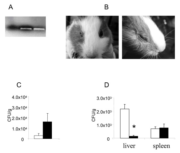
Figure 7.
Infection in guinea pigs caused by L. monocytogenes-infected T. pyriformis cysts. A. qPCR products resolved on 2,5 % agarose. 1 - negative control, 2 - L. monocytogenes culture lysates, 3 - lysates of T. pyriformis cysts infected with L. monocytogenes. B. L. monocytogenes associated conjunctivitis. On the left, conjunctivitis of the right eye caused by L. monocytogenes, the left eye was not infected; on the right, conjunctivitis caused by T. pyriformis cysts carrying L. monocytogenes. C. L. monocytogenes isolated from faeces of animals infected orally with L. monocytogenes (while columns) or with L. monocytogenes-infected cysts (black columns). D - bacterial loads in the liver and the spleen of animals infected orally with L. monocytogenes (while columns) or with L. monocytogenes-infected cysts (black columns) after 72 h post-infection. Data were expressed as the mean ± SE for groups of three animals. X, only one animal gave feces after 24 h. * p < 0,05

