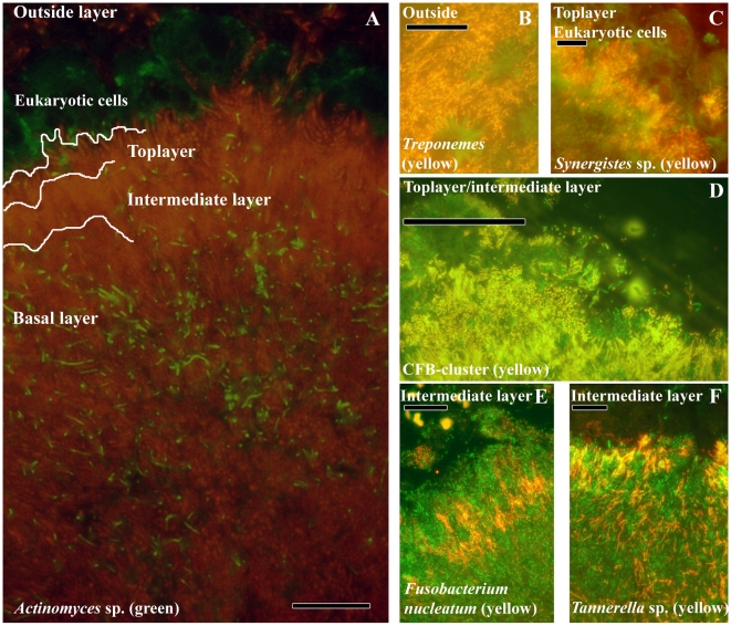
Figure 2. Localization of the most abundant species in subgingival biofilms.
(A) Overview of the subgingival biofilm with Actinomyces sp. (green bacteria), bacteria (red) and eukaryotic cells (large green cells on top). (B) Spirochaetes (yellow) outside the biofilm. (C) Detail of Synergistetes (yellow) in the top layer in close proximity to eukaryotic cells (green). (D) CFB-cluster (yellow) in the top and intermediate layer. (E) F. nucleatum in the intermediate layer. (F) Tannerella sp. (yellow) in the intermediate layer. Each panel is double-stained with probe EUB338 labeled with FITC or Cy3. The yellow color results from the simultaneous staning with FITC and Cy3 labeled probes. Bars are 10 µm.

