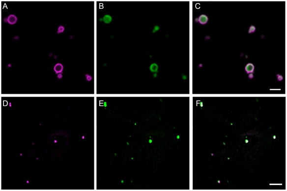
Figure 1. Localisation of PEX11e to peroxisomes.
eYFP-PEX11e (panel A, magenta) was transiently co-expressed with the peroxisomal marker CFP-SKL (panel B, green) in tobacco epidermal cells. The two fluorescent markers co-localise in small motile structures 1–2 micrometers in size, typical of peroxisomes (panel C). Arabidopsis suspension cells were immuno-labelled with anti-PEX11 antibody (D, magenta) and anti isocitrate lyase antibody (E, green) a peroxisomal (glyoxysomal) marker protein. The merged figure (F) shows PEX11 in punctate structures containing the glyoxysomal enzyme ICL. Scale bar 2 µm.

