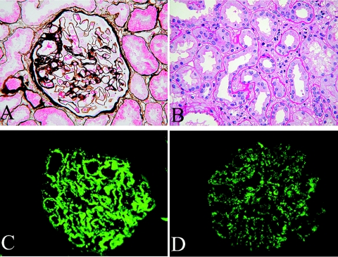
Figure 1.
Pathologic findings in mercury-induced MN. (A) Thickened glomerular basement membrane, as well as subepithelial fuchsinophilic deposits along the epithelium. Patient no. 2 (PASM staining; magnification, ×400). (B) Loss of the tubular brush border and focal concentration of interstitial infiltrating cells. Patient no. 10 (periodic acid-Schiff staining; magnification, ×400). (C) Deposits of IgG1 in the glomerular basement membrane (2+). Patient no. 2 (immunofluorescence staining; magnification, ×400). (D) Deposits of IgG4 in the glomerular basement membrane (1+). Patient no. 2 (immunofluorescence staining; magnification, ×400).

