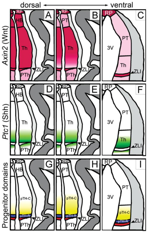
Fig. 3.
Schematic summary of Axin2 expression in relation to Shh activity and thalamic progenitor domains. Midline is to the left. A–C: Axin2 expression shown in pink. A: On dorsal sections, Axin2 is uniformly high in the thalamus as well as the ZLI, habenula (HB) and the roof plate (RP). The prethalamus (PTh) shows lower expression, which is shown in lighter pink. B: At the middle level, Axin2 expression is strong in the roof plate, the caudal part of the thalamus and the ZLI. C: The pretectum (PT), the rostral part of the thalamus just caudal to the ZLI and the prethalamus show weaker expression. At the most ventral level, Axin2 is strong only in the roof plate and the ZLI, whereas other part is uniformly weak. Not shown here is that the lateral edge of the thalamic ventricular zone shows strong Axin2 expression even at this ventral level (Fig. 3I, double arrowheads). D–F: Ptc1 expression shown in green to indicate the differential Shh signaling. On all three section planes, Ptc1 shows a rostral-high, caudal-low gradient of expression, whereas the ZLI does not express Ptc1. Ptc1 is also expressed in the most caudal part of the prethalamus. G–I: Thalamic progenitor domains. The thalamic ventricular zone, which lies caudal to the ZLI (red), is divided into two domains, pTH-R (blue) and pTH-C (yellow-white). pTH-R expresses transcription factors such as Ascl1 and Nkx2.2, whereas pTH-C expresses Neurog2 (=Neurogenin 2 or Ngn2), Olig2, and Dbx1. Olig2 is expressed in rostroventral-high, caudodorsal-low pattern (shown in the graded yellow color) and Dbx1 is expressed in the opposite pattern within pTH-C.

