
Fig. 6.
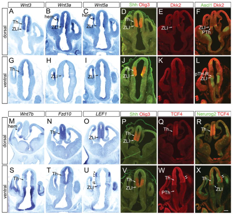
Differential expression of Wnt ligands and signaling components in thalamic progenitor cells at embryonic day (E) 11.5. Frontal sections E11.5 forebrain. A–X: A–F and M–R are sections that include dorsal part of the thalamus, whereas sections G–L and S–X contain more ventral part. A–D,G–J,M–P,S–V: In situ hybridization for Wnt3 (A,G), Wnt3a (B,H), Wnt5a (C,I), Wnt7b (M,S), Fzd10 (N,T), and LEF1 (O,U) are shown in comparison with double immunohistochemistry of Olig3 and Shh (D,J,P,V) that provides reference for the thalamic ventricular zone. E,F,K,L,Q,R,W,X: Also shown are immunohistochemistry for Dkk2 (E,K,F,L) and TCF4 (Q,R,W,X) in combination with Ascl1 (F,L) and Neurog2 (R,X). See text for more details. Th, thalamus; ZLI, zona limitans intrathalamica; PTh, prethalamus. Scale bar = 200 μm.