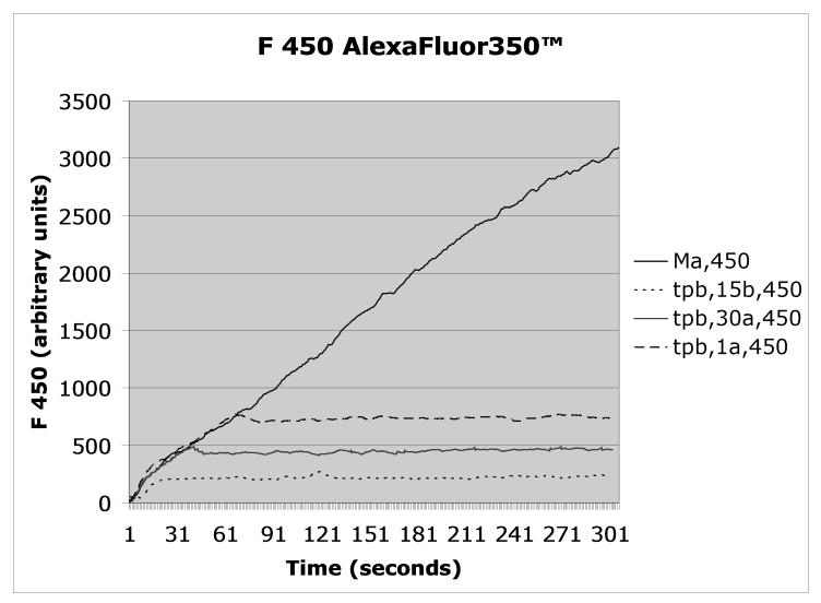
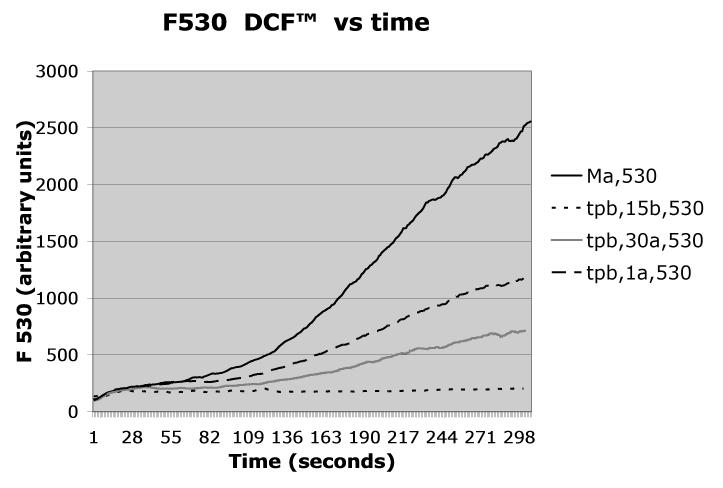
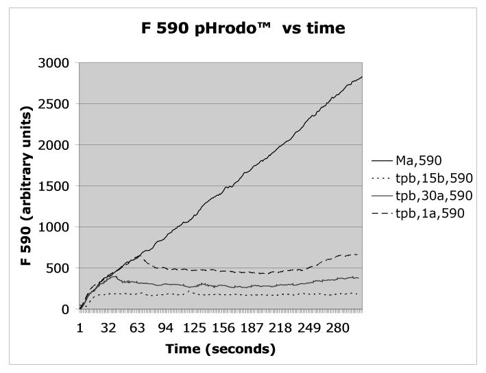
Figure 2.
Mycobacterium avium was triple labeled with AlexaFluor350™/pHrodo™/DCF™ according to Protocol 1, opsonized with autologous serum, and added to PMN at t=0 according to Protocol 2. Fluorescence emission was recorded continuously at 450nm (Alexafluor 350), 530 nm (DCF), or 590nm (pHrodo), a shown in the respective panels. As indicated in the figures’ legends, trypan blue (tpb) was added according to Protocol 3 to distinguish between enclosed (non-quenched) and bound but not enclosed (exposed to trypan blue, therefore quenched) organisms either 15 seconds before (15b) or at the indicated times after the Mycobacterium avium suspension (one second after = 1a, 30 seconds after = 30a). The control, i.e., unquenched, fluorescence is indicated as Ma.


