
Figure 1.
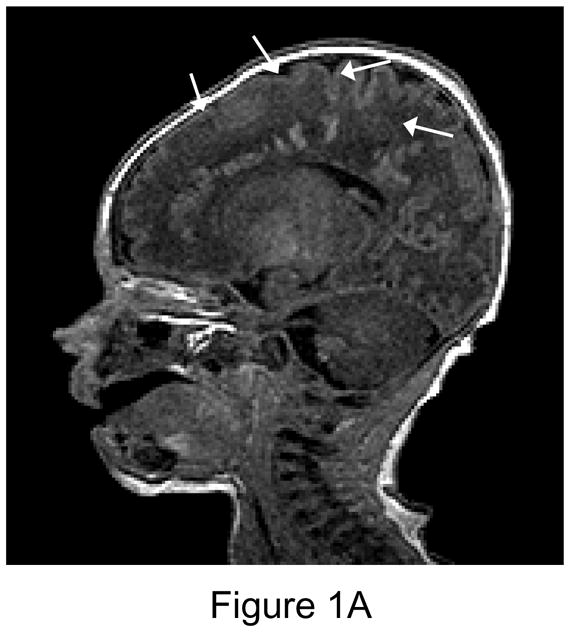
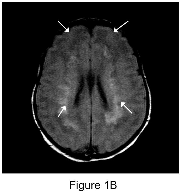
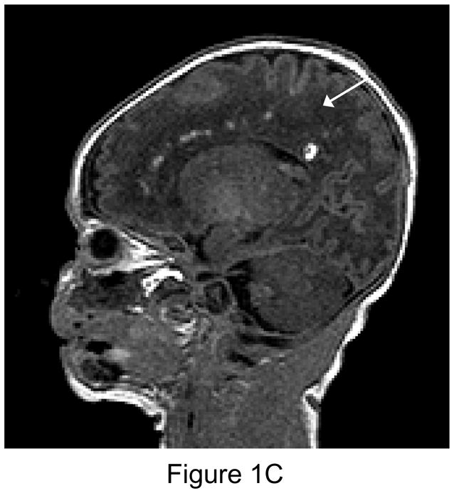
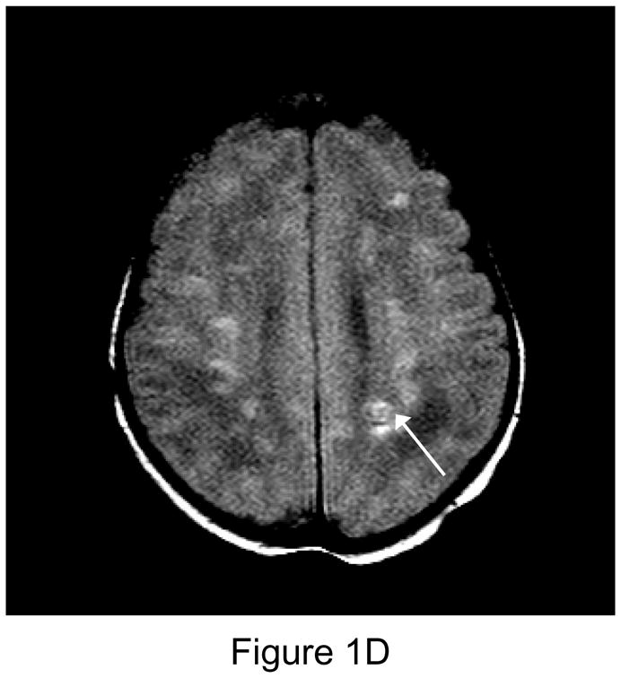
1A. Preoperative sagittal T1-weighted MR image of a 35 week gestational age infant with hypoplastic left heart syndrome. Extensive white matter injury (WMI) is present in the periventricular areas. (arrows). 1B. Preoperative axial proton-density T2 weighted image. Again note extensive WMI (arrows). 1C. 7-day postoperative T1 sagittal MRI after Norwood Stage I palliation. Note new intraparenchymal/intraventricular hemorrhage and infarction in the left paritrigonal region (arrow). 1D. Proton density T2-weighted image. Again note WMI and new hemorrhage (arrow). This patient had the single highest injury score on both preoperative, and postoperative MRI injury scale, at 11 points preoperatively, and 21 points postoperatively. (Refer to MRI Scoring Table in Appendix).