Fig. 9.
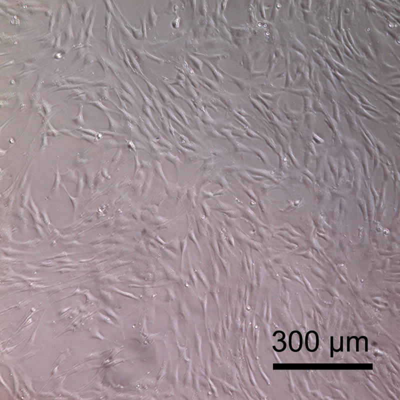
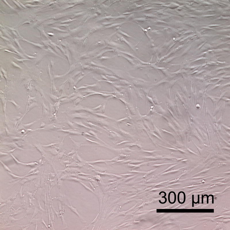
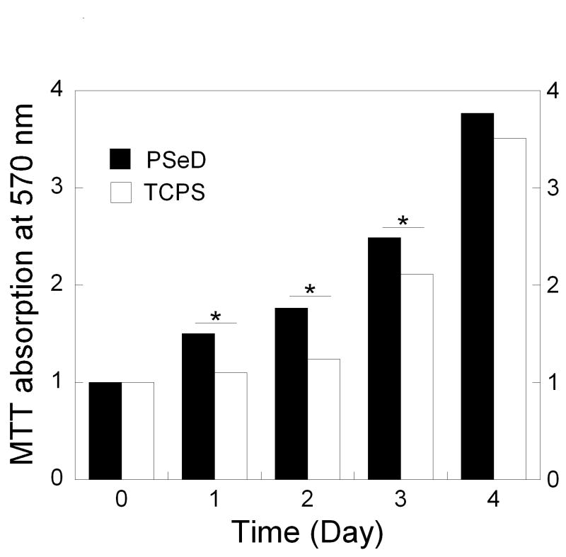
Baboon smooth muscle cells cultured on PSeD surface exhibited normal morphology, similar to that on TCPS. Representative phase contrast photomicrographs of baboon smooth muscle cells at day 3 after seeding in PSeD-coated wells (A) and TCPS wells (B) (magnification 100×, scale bar = 300 μm). (C) MTT assay of baboon smooth muscle cells in PSeD wells and TCPS wells showed that there were more metabolically active cells on PSeD than on TCPS in the first three days. Data represent mean ± SD. * Statistical significance (p < 0.05). Normalized values shown.


