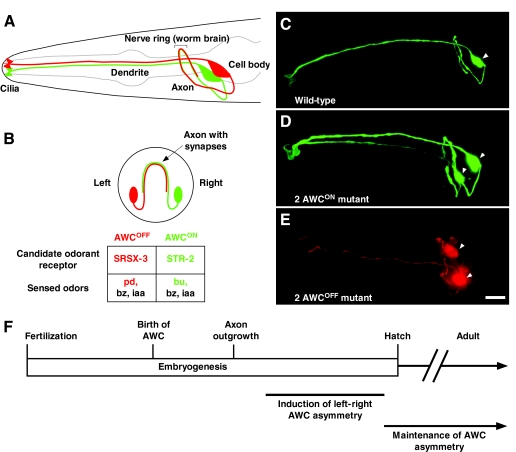
Fig. 1.
Left-right AWC neuronal asymmetry in C. elegans. (A,B) Amphid wing `C' (AWC) cell anatomy. Lateral (A) and transverse (B) views of a wild-type C. elegans head showing two bilateral AWC neurons. One AWC cell that expresses a GFP-tagged transgene of the candidate odorant receptor gene str-2 (str-2::GFP) (AWCON; right) is shown in green; the other AWC, with no str-2::GFP expression (AWCOFF; left), is shown in red. Within a population of worms, the left-right AWC asymmetry is stochastic, meaning that 50% of the animals display str-2 expression on the right, whereas the other 50% display str-2 expression on the left. The AWCON cell expresses the STR-2 candidate odorant receptor and senses butanone (bu), benzaldehyde (bz) and isoamyl alcohol (iaa). The AWCOFF cell expresses the SRSX-3 candidate odorant receptor and senses pentanedione (pd), bz and iaa (Bargmann et al., 1993; Wes and Bargmann, 2001). (C-E) Projections of micrograph stacks of wild-type and mutant animals expressing str-2::GFP (pseudo-colored green for GFP, red for DsRed). (C) Wild-type animals express str-2::GFP in one AWC neuron. (D) 2 AWCON animals express str-2::GFP in both AWC neurons. (E) Bilateral expression of the AWC marker transgene odr-1::DsRed in a 2 AWCOFF mutant showing the presence of both AWCs without str-2::GFP expression. (F) Time-line of developmental events in left-right AWC asymmetry. The AWC neurons are born at 300 minutes and their axons extend at 450 minutes after fertilization. AWC asymmetry is established during late embryogenesis and is maintained throughout the life of the animal. Arrowheads indicate the cell body of AWC neurons. Ventral is at the bottom in A-E, and anterior is to the left in A,C-E. Scale bar: 10 μm.

