Fig. 4.
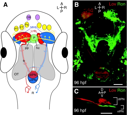
The asymmetric zebrafish habenular nuclei are central to the dorsal diencephalic conduction pathway. (A) The dorsal diencephalic conduction system. The ratio of lateral (red) to medial (blue) subnuclei is greater in the left habenula, but both sides receive input from both ipsilateral and contralateral neurons of the pallium, the eminentia thalami and the posterior tuberculum. The parapineal only sends projections to the left habenula, whereas the right habenula receives additional afferents from both olfactory bulbs. Habenular efferents course ventrally to the optic tecta in converging bundles known as the fasciculus retroflexi. Axons originating in the medial subnuclei innervate the ventral region of the interpeduncular nucleus, whereas those of lateral origin can target the dorsal interpeduncular nucleus. Some habenular axons also contact the raphe, just posterior to the interpeduncular nucleus. (B) Dorsal perspective confocal z-projection of Leftover (Lov)-positive (Lov+; red) and Right on (Ron)-positive (Ron+; green) neurons coursing from the habenular nuclei to the interpeduncular nucleus via the fasciculus retroflexi. (C) Lateral view of Lov+ and Ron+ axons, segregated into the dorsal and ventral domains of the interpeduncular nucleus. dIPN, dorsal interpeduncular nucleus; ET, eminentia thalami; fr, fasciculus retroflexus; hc, habenular commissure; LHb, lateral habenular subnuclei; MHb, medial habenular subnuclei; OB, olfactory bulb; OT, optic tectum; P, pineal organ; Pa, pallium; pp, parapineal organ; PT, posterior tuberculum; R, raphe nucleus; vIPN, ventral interpeduncular nucleus. Scale bars: 50 μm.

